Choosing the best fixation pin in trauma care is rarely a simple product comparison. In 2026, surgeons may consider Steinmann pins, Schanz pins, Kirschner wires, threaded pins, and hydroxyapatite-coated options. Each design serves a different mechanical and clinical purpose.
How to select fixation pins for trauma cases depends on fracture location, bone quality, soft-tissue damage, and the planned fixation frame. A thick Schanz pin may provide strong purchase in the tibia, while a smaller K-wire can support delicate fragments near the wrist. In osteoporotic bone, thread profile and holding strength become especially important. Pin diameter also matters. Too small may bend or loosen; too large can weaken the bone.
Clinical judgment remains central.
Experienced trauma teams assess radiographs, computed tomography findings, wound condition, and nearby nerves or vessels before choosing a pin. They also consider insertion stability, heat generation, pin-tract care, and later removal. Coated pins may reduce loosening in selected situations, but they are not a universal solution. Evidence is still uneven across patient groups and fixation systems.
The “best” pin is therefore the one that matches the injury, anatomy, and treatment plan. It should support alignment without creating unnecessary soft-tissue damage. Even careful choices can fail when swelling changes, infection develops, or bone quality is underestimated. This guide examines current fixation pin types, practical selection factors, and the limitations that clinicians should openly recognize.
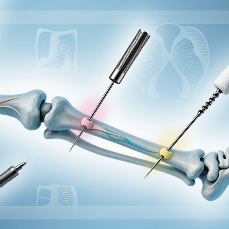
2026 Best Fixation Pin Types for Trauma Cases?
Understanding Fixation Pin Selection in Trauma Care
Choosing a fixation pin is not simply a hardware decision. It begins with fracture stability, bone quality, soft-tissue damage, and the treatment plan. Half-pins can provide strong purchase in external frames, especially when the main fragment offers enough safe bone. Smooth pins or wires may support smaller fragments, but they require careful tension and positioning. Threaded pins can improve grip, although excessive diameter or insertion force may weaken bone.
Pin selection should follow imaging, anatomical landmarks, and local surgical protocols. A pin must avoid nerves, vessels, joints, and future surgical pathways. In contaminated wounds, temporary fixation may protect alignment while soft tissues recover. The strongest-looking option is not always the safest. That point is easy to miss.
Tips: Match pin diameter to bone size and quality. Use fluoroscopy when anatomy is uncertain. Keep pin spacing adequate for frame stability. Monitor drainage, redness, loosening, and increasing pain. Pin-site care should follow the treating team’s protocol, not internet advice. Reassess fixation after swelling changes, because the initial frame may become less reliable. A useful reflection is this: did the pin improve control, or did it only add metal?
Fixation pin selection depends on fracture size, bone quality, soft-tissue damage, and treatment timing. Kirschner wires suit small fragments, hand fractures, and temporary control of alignment. Their slim profile causes less disruption, but they provide limited stability in heavy loading. Steinmann pins are thicker and useful for skeletal traction or temporary stabilization of larger bone segments.
Schanz half-pins commonly support external fixation frames in open fractures, polytrauma, and severe swelling. Threaded pins can improve purchase, especially in weaker bone, yet excessive diameter may create stress concentration. Pin placement must respect safe corridors, nearby nerves, vessels, and future surgical approaches. Imaging helps, but it does not replace careful anatomical judgment. No pin type wins every case.
Tips: Match pin diameter to the bone, not only the fracture pattern. Pre-drill when appropriate, avoid repeated passes, and monitor heat during insertion. Keep pins away from planned incisions. Check alignment, joint motion, skin tension, and distal circulation after fixation. A pin-site care plan should be practical for the patient and staff. Small errors matter. Teams should also question whether a temporary construct is becoming definitive without adequate reassessment.
Pin selection should follow anatomy, not habit. A thin smooth Kirschner wire suits small fragments, pediatric metaphyseal injuries, and temporary reduction. Its narrow diameter limits bone damage, but it may bend or migrate under heavy loading. Threaded pins can improve purchase in cancellous bone, especially near the distal radius or proximal tibia. They still require careful insertion to avoid joint penetration.
Fracture severity changes the design choice. Stable, low-energy fractures may tolerate percutaneous pins with external protection. Comminuted, open, or highly unstable injuries often need larger Schanz or Steinmann pins within an external frame. The 2019 Global Burden of Disease analysis estimated 178 million new fractures worldwide, showing why adaptable fixation strategies matter. Location remains decisive. Dense diaphyseal cortex favors strong bicortical purchase, while thin periarticular bone demands shorter, accurately placed pins.
The neat rule breaks.
Clinical guidance from major orthopedic trauma references stresses imaging, soft-tissue assessment, and repeated stability checks. A 2023 National Trauma Data Bank report also reflects the complex injury patterns seen in modern trauma care. Pin diameter, thread position, insertion angle, and frame distance can matter as much as the pin category. I would not treat any chart as absolute; swelling changes, bone quality is unpredictable, and an apparently stable construct may loosen. Surgeons should match the implant to the fracture, patient, and evolving wound condition.
Choosing fixation pins for trauma cases requires more than comparing catalog measurements. Material affects stiffness, corrosion resistance, imaging compatibility, and handling. Stainless steel remains familiar and mechanically dependable. Titanium is lighter and may create fewer imaging artifacts, but its flexibility can change construct behavior. The surgeon must match material properties with fracture pattern, bone quality, and planned loading.
Coatings deserve careful evaluation. A porous or roughened surface may improve bone contact in selected situations. However, it can also complicate removal or increase debris if the pin becomes damaged. Antimicrobial surfaces may appear attractive, yet their value depends on coating durability, local protocols, and infection risk. No coating replaces sterile technique and consistent pin-site care. Small details matter.
Pin size should support stability without creating unnecessary bone damage. Larger pins can improve bending resistance, especially in poor-quality bone. They may also increase thermal injury and weaken the cortex if inserted carelessly. Smaller pins reduce disruption, but they can fail under excessive load. Thread design, working length, insertion speed, and frame geometry matter as much as diameter. Fit matters.
In practice, pin selection is rarely perfect. Patient swelling can change access. Follow-up imaging may reveal unexpected loosening. A technically strong construct may still perform poorly when soft-tissue conditions are ignored. Reviewing outcomes, documenting complications, and adjusting protocols provide more reliable guidance than relying on material labels alone.
Choosing a fixation pin for trauma care depends on fracture pattern, bone quality, soft-tissue damage, and the required stability. Smooth pins may support temporary alignment, while threaded pins can improve resistance to migration. The choice should follow imaging, examination findings, and the surgeon’s treatment plan. A suitable pin cannot compensate for unsafe placement.
Safe placement begins with protecting nerves, vessels, tendons, and growth plates. The entry point should be planned using anatomical landmarks and appropriate imaging. Drilling should be controlled, with attention to heat, depth, and trajectory. Pins should not cross damaged skin unnecessarily.
Small details matter.
A misplaced pin can create pain, weakness, infection, or another operation.
Postoperative management requires clear instructions and regular review. The wound should remain clean and dry as directed, while redness, drainage, fever, increasing pain, numbness, or loose hardware needs prompt assessment. Pin sites should not be adjusted at home. Weight-bearing and movement limits must match the fixation strength and healing stage.
Follow-up imaging can reveal migration before symptoms become severe.
In practice, complications are not always preventable, and I would not promise otherwise. Even a technically sound procedure may face swelling, delayed healing, or patient-specific risks. Careful documentation, patient education, and honest reassessment remain essential.
Thin wires suit small bone fragments, hand fractures, and temporary alignment control. They disturb less tissue. However, they offer limited stability under heavy loading.
Thicker pins may support skeletal traction or larger bone segments. They can resist bending better. Their size may increase bone damage if inserted carelessly.
Half-pins commonly support frames during open fractures, severe swelling, or multiple injuries. They help stabilize limbs while soft tissues recover. Temporary fixation can become permanent unintentionally.
Match diameter to bone size, bone quality, and expected loading. Do not rely only on the fracture pattern. Large pins may improve strength but increase heat and cortical stress.
Threaded designs may improve purchase in weaker bone. Excessive diameter can create stress concentration. The strongest-looking option is not always safest.
Stainless steel offers familiar stiffness and dependable handling. Titanium is lighter and may reduce imaging artifacts. Its flexibility can change frame behavior. Material labels do not decide everything.
No. Rough or porous surfaces may improve bone contact in selected cases. They can complicate removal or create debris when damaged. Coatings never replace sterile technique or consistent site care.
Check alignment, joint movement, skin tension, and circulation beyond the fixation. Review imaging and nearby nerve or vessel safety. Watch for loosening, heat injury, swelling, or infection. Small errors matter.
Selecting the right fixation pin is essential for achieving stable fracture alignment, protecting soft tissue, and supporting efficient healing in trauma care. How to select fixation pins for trauma cases depends on the fracture pattern, anatomical location, injury severity, bone quality, and the required level of stability. Common options include smooth, threaded, half, and full pins, each offering different advantages for temporary or definitive fixation. Pin diameter, length, thread design, and insertion method should be chosen according to the patient’s anatomy and the mechanical demands of the injury.
Material and surface characteristics also influence performance, corrosion resistance, and tissue response. Appropriate placement should avoid nerves, vessels, joints, and damaged skin while maintaining secure purchase in healthy bone. After surgery, careful pin-site cleaning, wound observation, imaging follow-up, and timely management of loosening or infection are important. A structured, patient-specific approach helps clinicians balance stability, safety, and postoperative recovery.

Clerivida Medical