Why patient-specific implants are important is becoming a central question in modern reconstructive surgery. Standard implants follow common anatomical measurements, but patients rarely have standard anatomy. Severe bone loss, previous surgery, trauma, or unusual joint geometry can make a close fit difficult. A patient-specific implant uses medical imaging, usually CT data, to guide digital planning and manufacturing. The result may better match the patient’s bone shape, surgical goals, and available fixation surfaces.
The American Joint Replacement Registry’s 2024 Annual Report analyzes millions of hip and knee procedures, showing the growing need for reliable long-term implant surveillance. Its data also reinforces an important point: implant success depends on more than design. Surgical technique, infection prevention, rehabilitation, and patient health remain decisive. The U.S. Food and Drug Administration’s guidance on additive manufacturing describes the need for strict material control, validated production, and device-specific testing. Personalization does not remove those responsibilities.
The practical value can be easy to visualize. A surgeon may rotate a three-dimensional model before entering the operating room. A custom implant may then fill a complex defect more precisely than an off-the-shelf option. That can support bone preservation and simplify difficult reconstruction. However, the evidence is still developing. Not every patient needs a custom device, and customization does not guarantee fewer complications. Cost, production time, imaging quality, and revision planning also matter. These limitations deserve honest discussion. The seven reasons explored here connect engineering precision with clinical judgment, patient safety, and measurable outcomes—not marketing promises.
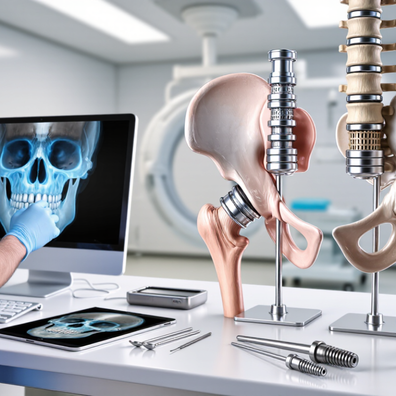
Patient-specific implants begin with trustworthy anatomy, not software alone. CT datasets commonly use 0.5–1 mm slice resolution for detailed bone assessment. This range helps capture thin structures, fracture edges, and irregular defects. It also supports more accurate segmentation and virtual surgical planning.
The FDA’s Technical Considerations for Additive Manufactured Medical Devices emphasizes controlled imaging, segmentation, and design verification. DICOM standards separately record slice thickness and image spacing, which prevents a frequent planning mistake. They are not always identical. A 0.5 mm slice does not automatically guarantee perfect three-dimensional detail.
Small errors can become clinically meaningful. A 1 mm boundary shift may affect screw clearance, implant seating, or bone contact. The AAPM Report No. 83 identifies spatial resolution, contrast, noise, and geometric accuracy as important CT quality factors. These variables interact. A thinner slice may increase noise and produce a less reliable contour. That assumption needs questioning. Patient movement, metal artifacts, reconstruction kernels, and calibration can also distort anatomy. Experienced teams therefore review source images, not only the exported model. They document scanner settings, orientation, and segmentation decisions. The process is highly precise, but never entirely automatic.
7 Best Reasons Why Patient Specific Implants Matter?
Reasons 1–2—Fit and Alignment: Planning Targets Submillimeter Accuracy
Patient-specific implants begin with the patient’s own CT anatomy, not an average template. Engineers map bone loss, joint surfaces, and safe screw corridors in three dimensions. The plan may target alignment within one millimeter. Small errors matter. A two-millimeter shift can alter contact pressure, limb mechanics, or soft-tissue tension. In complex reconstruction, precise fit can also reduce intraoperative reshaping and unnecessary bone removal.
Large registry data supports this need for disciplined planning. The National Joint Registry’s 2024 report includes more than three million hip and knee procedures. It shows that revision risk varies with patient factors, implant choice, and surgical indication. The data does not prove that every patient-specific implant prevents revision. It does show why alignment cannot rely on visual estimation alone. Digital planning creates measurable targets before the patient enters the operating room.
However, a submillimeter plan is not a submillimeter result. Registration errors, saw movement, implant seating, and soft-tissue forces can change the final position. Our planning assumptions can still be wrong. That weakness deserves attention. Surgeons should compare the virtual plan with intraoperative landmarks and postoperative imaging. A 2023 review in EFORT Open Reviews also emphasized that patient-specific technologies require careful validation, not automatic trust. Fit is valuable only when anatomy, surgical execution, and verification agree.
Patient-specific implants matter because bone is rarely symmetrical or predictable. A three-dimensional model can reflect a patient’s defect, anatomy, and available bone more closely than a standard shape. That closer fit may improve contact and help surgeons plan fixation before entering the operating room. Still, imaging is not perfect. Small errors in scans or manufacturing can affect the final result.
Bone response is another important reason. Carefully engineered pores measuring about 300–800 μm can provide space for vascular tissue and new bone growth. This structure may support osseointegration, allowing bone to grow into the implant surface over time. The pore range is useful, not magical. Material choice, pore connectivity, surface quality, loading, and the patient’s biology also influence healing.
Fixation depends on both design and surgical execution. A patient-specific implant can offer planned screw paths, better contact areas, and more stable load transfer around weakened bone. Stability matters because excessive micromotion may interfere with bone attachment. However, an accurate design cannot replace careful preparation or sound clinical judgment. Smoking, infection risk, poor bone quality, and incomplete rehabilitation may still compromise integration. Surgeons must review each case individually, question the digital plan, and adjust it when real anatomy differs from the model. That practical flexibility is easy to overlook.
Patient-specific implants matter most when workflow cannot tolerate guesswork. Three-dimensional planning can show bone loss, fixation zones, and implant position before the patient enters the operating room. In complex reconstruction, that preparation may reduce instrument changes and intraoperative improvisation. However, the evidence is not tidy. A 2020 meta-analysis of 26 randomized trials found no consistent reduction in operating time with patient-specific instrumentation for knee replacement. The gain is not guaranteed.
OR minutes still have practical value. A shorter procedure can reduce anesthesia exposure, staff burden, and room occupancy. Yet planning, imaging, and manufacturing add time before surgery. That trade-off deserves measurement. Hospitals should compare total pathway time, not only incision-to-closure minutes. Their local results may differ.
Recovery requires the same discipline. The American College of Surgeons National Surgical Quality Improvement Program tracks 30-day complications, readmissions, reoperations, and mortality. These measures offer a useful benchmark for patient-specific implant programs. Published reconstruction studies often report small cohorts, so impressive early results may not generalize. A 30-day review should examine wound problems, unplanned returns, pain-related visits, and mobility milestones. Better alignment may support recovery, but it does not replace rehabilitation or careful patient selection. Some outcomes remain uncertain. That uncertainty should stay visible.
| Workflow or outcome measure | Conventional, surgeon-shaped implant | Patient-specific implant | Practical interpretation |
|---|---|---|---|
| Preoperative planning | Implant selection and intraoperative shaping may be finalized during the surgical episode. | Requires CT-based planning, design approval, manufacturing, sterilization, and delivery before surgery. | Patient-specific planning can make the operation more predictable, but it adds lead time before the procedure. |
| Intraoperative shaping and trial fitting | More manual contouring, repeated trial fitting, and adjustment may be required, particularly in anatomically complex defects. | The implant is designed to match the planned defect, which may reduce intraoperative contouring and fitting steps. | The greatest workflow benefit is generally expected in irregular, asymmetric, or difficult-to-access defects. |
| Operating-room time | May be prolonged when shaping, repositioning, or repeated fitting is necessary. | May reduce implant-fitting and contouring time in selected cases; the total operation may still be dominated by exposure, reconstruction, closure, and anesthesia. | Comparative studies do not establish one universal time saving. The effect depends on anatomy, surgical technique, implant type, and team experience. |
| Need for intraoperative modification | More likely when the defect is complex or the preoperative estimate does not match the exposed anatomy. | Usually less contour modification is anticipated, although intraoperative adjustment can still be necessary. | Patient-specific design improves fit planning, but it does not eliminate surgical judgment or unexpected findings. |
| Early postoperative imaging | Position and contour are assessed against the surgeon’s intraoperative reconstruction plan. | Position and contour can be assessed against the approved digital design and planned anatomy. | Digital planning provides an additional reference for evaluating reconstruction accuracy; it does not replace clinical assessment. |
| Length of hospital stay | Varies mainly with procedure complexity, comorbidities, pain control, mobility, and complications. | May be shorter if the implant improves operative efficiency, but a consistent independent reduction has not been established across procedures. | Hospital stay should not be predicted from implant design alone. |
| 30-day reoperation | Risk is influenced by infection, wound problems, malposition, exposure, hematoma, and the underlying disease. | Patient-specific fit may reduce technical mismatch, but available comparative evidence does not demonstrate a universal reduction in 30-day reoperation. | A lower reoperation rate should be considered a possible benefit in selected cases, not a guaranteed outcome. |
| 30-day surgical-site infection | Risk is primarily related to contamination, soft-tissue condition, operative duration, patient factors, and procedure type. | A customized shape does not by itself remove infection risk; evidence does not support a consistent implant-design effect across all indications. | Infection prevention depends more broadly on surgical and perioperative practice than on customization alone. |
| 30-day wound or implant exposure | Can occur when soft-tissue coverage is inadequate or healing is impaired. | Improved contour may support reconstruction in selected anatomy, but soft-tissue quality remains the primary determinant. | Customization may assist fit, but it cannot compensate for inadequate coverage or poor healing capacity. |
| 30-day patient recovery | Recovery depends on the same clinical factors and may be affected by longer or more technically demanding surgery. | A smoother workflow may support recovery when it meaningfully reduces operative burden, but patient-reported recovery advantages remain procedure-specific. | The strongest evidence-based claim is improved planning and potential workflow efficiency—not a guaranteed improvement in every 30-day outcome. |
Evidence note: Operating-room time and 30-day outcomes vary substantially by anatomical site, reconstruction complexity, patient risk, surgical team, and implant material. Published comparative evidence supports potential workflow advantages for patient-specific implants in selected cases, but it does not establish a universal reduction in operating time, complications, hospital stay, or 30-day reoperation.
Reason 7—Long-Term Value: Audit Revisions, Complications, and 5-Year Survival
Patient-specific implants matter because their value extends beyond the operating room. Their performance should be reviewed through revisions, complications, function, and five-year survival. A smooth recovery is encouraging, but it does not prove durable success. Long-term follow-up reveals loosening, infection, alignment changes, and unexpected bone loss.
Surgeons and hospitals should maintain clear records before and after implantation. These records can include imaging, rehabilitation progress, pain scores, implant position, and secondary procedures. Auditing this information supports better clinical decisions and more honest patient discussions. Published studies and independent registries are useful, but results may differ between hospitals. Surgical technique, patient health, implant design, and follow-up quality all influence outcomes. Five-year survival is important, yet it should never be treated as a guarantee.
Tips: Ask how long-term outcomes are measured. Check whether complications include minor events and revision surgery. Request evidence from comparable patients, not only laboratory testing. Review the plan for future monitoring. A practical detail matters: patients often lose follow-up after feeling better. That gap can hide problems.
Patient-specific planning may reduce technical mismatch, but it cannot remove biological risk. My own assumption would need testing against larger, longer datasets. Some early improvements may fade. That is why durable value depends on transparent audits, experienced clinical judgment, and continued patient monitoring.
Published clinical series and systematic reviews of patient-specific 3D-printed pelvic and acetabular implants report heterogeneous outcomes. The ranges below show commonly reported values used when auditing complications, revisions, and long-term implant survival.
Data shown as reported literature ranges rather than a pooled estimate. Outcomes vary with anatomy, reconstruction complexity, follow-up duration, tumor or revision status, surgical technique, and patient risk profile. A five-year survival of 85–95% means that approximately 85–95% of implants remained unrevised at five years in the reported cohorts.
Evidence context: peer-reviewed systematic reviews and clinical case series of custom and patient-specific pelvic or acetabular implants; small cohorts and inconsistent follow-up remain important limitations.
CT scans commonly use 0.5–1 mm slice resolution. This helps show thin bone, fracture edges, and irregular defects. Finer slices are not automatically better.
They describe different parts of the dataset. A 0.5 mm slice may still have different spacing between images. Confusing them can distort three-dimensional planning. Small mistakes matter.
No. Patient movement, metal artifacts, noise, and calibration can distort anatomy. Reconstruction settings also affect the visible bone boundary. Experienced teams should review original images and exported models.
Three-dimensional planning can identify bone loss, fixation zones, and implant position before surgery. It may reduce instrument changes and improvisation. The benefit is not guaranteed.
No. Research has not shown consistent time savings in every procedure. Planning and manufacturing also require extra time. Hospitals should measure the entire pathway, not only operating room minutes.
A 30-day review can include wound problems, readmissions, reoperations, pain-related visits, and mobility changes. Unplanned returns also matter. Better alignment does not replace rehabilitation.
Teams should track complications, revisions, function, imaging, pain, and implant position. Five-year survival is useful, but it is not a promise. Some early improvements may fade.
Ask how complications and revisions are recorded. Request evidence from similar patients, not laboratory testing alone. Ask who reviews future scans and when. Feeling better can hide a problem.
Why patient-specific implants are important lies in their ability to connect anatomical data with individualized surgical planning. Using CT scans commonly reconstructed at 0.5–1 mm slice resolution, clinicians can design implants and surgical guides that closely match a patient’s bone structure. This supports planning targets at submillimeter accuracy, which may improve fit, alignment, load distribution, and procedural predictability compared with generalized solutions.
Patient-specific design can also incorporate fixation features such as 300–800 μm porous structures, which may encourage bone ingrowth and stable osseointegration when clinically appropriate. Beyond the implant itself, the approach may streamline operating-room workflow, reduce unnecessary adjustments, and support smoother recovery. Its value should be assessed through measurable evidence, including operating time, 30-day outcomes, revision rates, complications, and five-year survival. By combining precise imaging, tailored engineering, and long-term clinical auditing, patient-specific implants offer a structured way to pursue safer, more durable, and more efficient care.

Clerivida Medical